Author's details
- Dr Abdulgafar Lekan Olawumi
- MBBS, MHE, MPH, FWACP, FMCFM
- Department of Family Medicine and Geriatric Centre of Aminu Kano Teaching Hospital Kano, Nigeria.
Reviewer's details
- Dr Kolade-Salawu Ayobami Nafisat
- (FMCFM)
- General Hospital Moniya, Ibadan.
- Date Uploaded: 2025-05-23
- Date Updated: 2025-12-22
Atopic Dermatitis
Key Messages
- Atopic dermatitis is a chronic, itchy inflammatory skin disease caused by barrier dysfunction, immune imbalance, genetics, and environmental triggers.
- It presents with dry, pruritic, eczematous lesions that vary by age and skin tone, and often coexists with other atopic conditions.
- The burden is rising in sub-Saharan Africa, with major quality-of-life impact and limited access to care.
- Diagnosis is clinical, and management centers on education, trigger avoidance, emollients, topical anti-inflammatories, and infection control.
Eczema, medically known as atopic dermatitis, is a chronic, relapsing inflammatory skin condition that poses a significant burden on affected individuals and healthcare systems worldwide. It is primarily characterized by intense pruritus (itching), erythema (redness), xerosis (dry skin), and the presence of eczematous lesions, which may evolve in appearance depending on the stage of the disease which may be acute, subacute, or chronic. In the acute phase, lesions are often erythematous and may exhibit vesiculation and oozing, while chronic eczema is typically associated with lichenification, scaling, and thickened skin due to repeated scratching.
Atopic dermatitis (AD) is strongly associated with a personal or family history of atopic diseases, such as asthma, allergic rhinitis, or food allergies, highlighting its genetic and immunologic basis. The condition is thought to result from a complex interplay of skin barrier dysfunction, immune dysregulation, genetic predisposition, and environmental triggers. These may include irritants, allergens, infections, stress, and climatic factors. Although eczema is most commonly diagnosed in early childhood, it can persist into adulthood or even present de novo in later life. The chronicity and recurrence of symptoms can significantly impair quality of life, leading to sleep disturbances, psychological distress, and increased susceptibility to secondary infections. Understanding epidemiology, pathophysiology, and clinical presentation of eczema is essential for effective diagnosis and management, particularly in resource-limited settings where misdiagnosis and under-treatment are common.
Pathophysiology of AD
AD is a chronic inflammatory skin condition resulting from a combination of skin barrier dysfunction, immune dysregulation, genetic predisposition, and environmental factors. Mutations in the filaggrin (FLG) gene and other skin barrier proteins impair the skin’s protective function, leading to increased trans-epidermal water loss and enhanced penetration of allergens, irritants, and microbes. This compromised barrier contributes to dry, sensitive skin and persistent inflammation. Environmental triggers such as climate extremes (heat, cold, humidity), irritants (soaps, detergents), allergens (dust mites, pollen, pet dander), and air pollution can aggravate symptoms. Additionally, lifestyle-related factors like urban living, poor hygiene, stress, and dietary changes have been associated with the rising prevalence of AD. Immunologically, AD is characterized by a dominant Th2 immune response in acute phases, involving cytokines like IL-4, IL-5, and IL-13, with progression to Th1, Th17, and Th22 responses in chronic stages. Colonization by Staphylococcus aureus and the itch-scratch cycle further exacerbate skin damage and inflammation, perpetuating the disease process.
Burden of AD in Sub-Saharan Africa
AD is an increasingly recognized public health concern in sub-Saharan Africa (SSA), with rising prevalence, particularly among children and urban populations. For instance, studies have reported varying prevalence rates: 1.6% in Ghana, 4.0% in Gabon, and 0.8% in Rwanda among schoolchildren aged 4 to 20 years.8 In Senegal, a prevalence of 12.2% was observed among children under 15 years, while a study in Nigeria reported a prevalence of 10% among children and adults.8,9 The burden of AD extends beyond physical symptoms like pruritus and skin infections; it significantly impacts psychosocial well-being, leading to reduced quality of life, school absenteeism, and stigma. Limited access to dermatological care, diagnostic tools, and appropriate treatments further complicates disease management in many SSA settings. Cultural perceptions of skin diseases may delay care-seeking and promote the use of unregulated traditional remedies, potentially worsening outcomes.
Risk factors
Several socioeconomic and environmental factors have been associated with the rising prevalence of atopic eczema globally, particularly in developing regions. Smaller family sizes are linked to reduced early childhood exposure to microbes, which may hinder immune system development, a concept known as the "hygiene hypothesis". Similarly, increased household income and higher levels of parental education are often associated with lifestyle and environmental changes, including the use of more household chemicals, processed foods, and lower microbial exposure. Furthermore, migration from rural to urban settings tends to increase exposure to air pollutants and allergens while decreasing contact with natural environments, both of which can contribute to the onset of AD. Another key factor is the increased use of antibiotics, especially early in life, which can disrupt the gut microbiome and immune regulation, making individuals more susceptible to allergic diseases such as atopic dermatitis.
Clinical features
A significant proportion of individuals with AD (about 50–70%) have a positive family history of atopic diseases, emphasizing the strong genetic predisposition associated with the condition. Additionally, around two-thirds of patients with AD also report a personal or family history of other atopic disorders, such as asthma, allergic rhinitis, or allergic conjunctivitis. This constellation of comorbidities further supports the concept of the "atopic march," in which different allergic conditions may develop sequentially over time in genetically predisposed individuals.
The clinical presentation of atopic dermatitis (AD) in sub-Saharan Africa (SSA) is broadly similar to global patterns but may show some distinct variations influenced by climate, skin type, and environmental exposures. Common features include intense pruritus (itching), dry and scaly skin (xerosis), erythema, and eczema lesions that may become lichenified due to chronic scratching. In acute stages, weeping or crusted lesions may be observed, especially with secondary bacterial infection, which is commonly caused by Staphylococcus aureus.
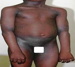
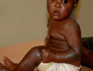
In dark skin tones, inflammation may appear violaceous or hyperpigmented rather than red, and post-inflammatory hyperpigmentation is often more pronounced and persistent. The distribution of lesions varies by age: infants typically present with facial and extensor involvement, while older children and adults show lesions in flexural areas such as the elbows, knees, neck, and wrists. Recurrent flares, sleep disturbance due to itching, and skin thickening are common in chronic cases.


Figure 1: Showing distribution of the atopic eczema rashes in an infant and adolescent.
Mode of diagnosis
In resource-limited settings, diagnosis is based on history and physical examination, using standardized clinical criteria. The most commonly used is “Hanifin and Rajka Criteria”, which requires: 3 or more major criteria, such as: pruritus, chronic or relapsing dermatitis, personal or family history of atopy, and typical distribution of skin lesions, with 3 or more minor criteria, such as: xerosis, ichthyosis, early onset, elevated serum IgE levels, chelitis, recurrent conjunctivitis, etc.
Differential diagnosis
Several skin conditions can mimic AD due to similarities in itching or rash patterns:
- Seborrheic Dermatitis - affects scalp, face (especially nasolabial folds), and chest, with greasy, yellowish scales rather than dry, eczematous patches.
- Contact Dermatitis - history of exposure to allergens or irritants is a clue. Rashes are often localized to the site of exposure.
- Scabies - commonly affect finger webs, wrists, waistline, and genitals. Burrows might be present and family members often affected.
- Psoriasis - well-demarcated, thick plaques with silvery scale. It is less pruritic than AD and typically not flexural.
- Tinea (Dermatophytosis) - annular (ring-like) lesions with central clearing. Positive KOH test can help confirm fungal infection.
- Ichthyosis Vulgaris - generalized dry, and scaly skin, which is typically hereditary and often co-exists with or mimics AD.9-11
Treatment
Treatment involves both pharmacological and non-pharmacological approaches.
Non-pharmacologic:
This approach is essential for enhancing patient satisfaction, improving adherence to treatment, and ensuring optimal treatment outcomes
- Patient education – educate patients and caregivers about the chronic and relapsing nature of AD. Clear communication on the importance of adherence to treatment, skincare routines, and identification of triggers is crucial.
- Avoidance of Triggers – identify and minimize exposure to known aggravating factors such as harsh soaps, detergents, fragrances, wool or rough fabrics, dust mites, pet dander, pollen, mold, emotional stress, overheating, and certain foods in sensitized individuals.
- Control of Exacerbating Environmental Factors – environmental modifications can help reduce atopic dermatitis flares. These include using air humidifiers in dry climates, wearing soft and breathable clothing, maintaining a cool and well-ventilated home, and minimizing exposure to cigarette smoke and indoor pollutants.
Pharmacologic:
- Restoration of Skin Barrier Function and Hydration: Daily application of emollients such as Vaseline, shea butter cream, other unscented petroleum jelly, is crucial to reduce trans-epidermal water loss and maintain skin integrity. They should be applied liberally and frequently, especially after bathing. Ointments or creams are preferred over lotions for better moisture retention. Continued use during symptom-free periods helps sustain remission and prevent flare-ups.
- Control of inflammation: Managing inflammation is essential during flare-ups of atopic dermatitis. Sedating antihistamines may help relieve nighttime itching, while topical anti-inflammatory therapies form the cornerstone of treatment.
- Topical Corticosteroids (TCS) - are the first-line option for acute inflammation, with potency tailored to the severity and location of the lesions.
- Topical Calcineurin Inhibitors (TCIs) such as tacrolimus and pimecrolimus are ideal for sensitive areas like the face, eyelids, and skin folds, and are effective for long-term maintenance to prevent relapses.
Treatment of Bacterial Superinfection: Secondary bacterial infections, commonly caused by Staphylococcus aureus, are frequent in atopic dermatitis. Topical or systemic antibiotics should be used when signs of infection, such as oozing, crusting, or increased redness are present. In recurrent cases, antiseptic measures like diluted bleach baths can help reduce bacterial colonization and prevent further flare-ups.
An interesting case we managed involved an infant with atopic dermatitis (AD) whose mother got distressed by persistent flare-ups despite home care, sought spiritual intervention, believing her child had been bewitched by her co-wife. This highlights a common challenge in resource-limited settings where chronic conditions like AD may be misunderstood due to cultural beliefs and limited awareness. Patient education in such cases is crucial. It helps caregivers understand the chronic and relapsing nature of AD, the role of genetic and environmental factors, and the importance of consistent skincare and medical treatment. By clearly explaining the disease process and demonstrating proper use of emollients and topical medications, healthcare providers can build trust, dispel myths, and prevent harmful delays in care. Educating caregivers empowers them to manage the condition confidently and reduces dependence on unverified remedies.
A 6-year-old child presents with dry, itchy, red patches on the face, elbows, and knees that have been present for several months. The child has a family history of asthma and allergic rhinitis. The rash worsens during the winter months and is aggravated by harsh soaps and scratching.
On examination, there are erythematous, excoriated lesions with areas of lichenification on the flexural surfaces. There are no signs of secondary infection.
A diagnosis of atopic dermatitis (atopic eczema) is made, linked to the patient’s family history of allergic conditions. Treatment includes moisturizers, avoiding irritants, and topical corticosteroids to control flare-ups. The child is also advised to use mild soaps and avoid scratching. Follow-up is scheduled to monitor the condition.
- Nemeth V, Syed HA, Evans J. Eczema. 2024. In: StatPearls [Internet]. Treasure Island (FL): StatPearls Publishing; 2025. PMID: 30855797.
- Sohn A, Frankel A, Patel RV, Goldenberg G. Eczema. Mt Sinai J Med. 2011;78(5):730-9. doi: 10.1002/msj.20289. PMID: 21913202.
- Schmid-Grendelmeier P, Takaoka R, Ahogo KC, Belachew WA, Brown SJ, Correia JC, et al. Position Statement on Atopic Dermatitis in Sub-Saharan Africa: current status and roadmap. J Eur Acad Dermatol Venereol. 2019;33(11):2019-2028. doi: 10.1111/jdv.15972.
- Al-Afif KAM, Buraik MA, Buddenkotte J, Mounir M, Gerber R, Ahmed HM, et al. Understanding the Burden of Atopic Dermatitis in Africa and the Middle East. Dermatol Ther (Heidelb). 2019;9(2):223-241. doi: 10.1007/s13555-019-0285-2.
- Kouotou EA, Nansseu JR, Minlo Nyangon EF, Tounouga DN, Defo D, Mendouga Menye CR, et al. Atopic dermatitis in adults from sub-Saharan Africa: epidemiological and clinical patterns, severity, and quality of life. Our Dermatol Online. 2021;12(1):1-8
- Bayonne-Kombo E, Loubove H, Voumbo Mavoungou Y, Gathsé A. Clinical Aspects of Atopic Dermatitis of Children in Brazzaville, Congo. Open Dermatol J 2019;13. http://dx.doi.org/10.2174/1874372201913010061
- Paul RE, Sakuntabhai A. Atopic dermatitis: the need for a sub-Saharan perspective. EMJ Allergy Immunol. 2016;1(1):58-64.
- Ibekwe PU, Ukonu BA. Prevalence of Atopic Dermatitis in Nigeria: A Systematic Review and Meta-analysis. Nigerian Journal of Dermatology 2021;11(2): 7-17.

More topics to explore
Author's details
Reviewer's details
Atopic Dermatitis
- Background
- Symptoms
- Clinical findings
- Differential diagnosis
- Investigations
- Treatment
- Follow-up
- Prevention and control
- Further readings
Eczema, medically known as atopic dermatitis, is a chronic, relapsing inflammatory skin condition that poses a significant burden on affected individuals and healthcare systems worldwide. It is primarily characterized by intense pruritus (itching), erythema (redness), xerosis (dry skin), and the presence of eczematous lesions, which may evolve in appearance depending on the stage of the disease which may be acute, subacute, or chronic. In the acute phase, lesions are often erythematous and may exhibit vesiculation and oozing, while chronic eczema is typically associated with lichenification, scaling, and thickened skin due to repeated scratching.
Atopic dermatitis (AD) is strongly associated with a personal or family history of atopic diseases, such as asthma, allergic rhinitis, or food allergies, highlighting its genetic and immunologic basis. The condition is thought to result from a complex interplay of skin barrier dysfunction, immune dysregulation, genetic predisposition, and environmental triggers. These may include irritants, allergens, infections, stress, and climatic factors. Although eczema is most commonly diagnosed in early childhood, it can persist into adulthood or even present de novo in later life. The chronicity and recurrence of symptoms can significantly impair quality of life, leading to sleep disturbances, psychological distress, and increased susceptibility to secondary infections. Understanding epidemiology, pathophysiology, and clinical presentation of eczema is essential for effective diagnosis and management, particularly in resource-limited settings where misdiagnosis and under-treatment are common.
- Nemeth V, Syed HA, Evans J. Eczema. 2024. In: StatPearls [Internet]. Treasure Island (FL): StatPearls Publishing; 2025. PMID: 30855797.
- Sohn A, Frankel A, Patel RV, Goldenberg G. Eczema. Mt Sinai J Med. 2011;78(5):730-9. doi: 10.1002/msj.20289. PMID: 21913202.
- Schmid-Grendelmeier P, Takaoka R, Ahogo KC, Belachew WA, Brown SJ, Correia JC, et al. Position Statement on Atopic Dermatitis in Sub-Saharan Africa: current status and roadmap. J Eur Acad Dermatol Venereol. 2019;33(11):2019-2028. doi: 10.1111/jdv.15972.
- Al-Afif KAM, Buraik MA, Buddenkotte J, Mounir M, Gerber R, Ahmed HM, et al. Understanding the Burden of Atopic Dermatitis in Africa and the Middle East. Dermatol Ther (Heidelb). 2019;9(2):223-241. doi: 10.1007/s13555-019-0285-2.
- Kouotou EA, Nansseu JR, Minlo Nyangon EF, Tounouga DN, Defo D, Mendouga Menye CR, et al. Atopic dermatitis in adults from sub-Saharan Africa: epidemiological and clinical patterns, severity, and quality of life. Our Dermatol Online. 2021;12(1):1-8
- Bayonne-Kombo E, Loubove H, Voumbo Mavoungou Y, Gathsé A. Clinical Aspects of Atopic Dermatitis of Children in Brazzaville, Congo. Open Dermatol J 2019;13. http://dx.doi.org/10.2174/1874372201913010061
- Paul RE, Sakuntabhai A. Atopic dermatitis: the need for a sub-Saharan perspective. EMJ Allergy Immunol. 2016;1(1):58-64.
- Ibekwe PU, Ukonu BA. Prevalence of Atopic Dermatitis in Nigeria: A Systematic Review and Meta-analysis. Nigerian Journal of Dermatology 2021;11(2): 7-17.

Content
Author's details
Reviewer's details
Atopic Dermatitis
Background
Eczema, medically known as atopic dermatitis, is a chronic, relapsing inflammatory skin condition that poses a significant burden on affected individuals and healthcare systems worldwide. It is primarily characterized by intense pruritus (itching), erythema (redness), xerosis (dry skin), and the presence of eczematous lesions, which may evolve in appearance depending on the stage of the disease which may be acute, subacute, or chronic. In the acute phase, lesions are often erythematous and may exhibit vesiculation and oozing, while chronic eczema is typically associated with lichenification, scaling, and thickened skin due to repeated scratching.
Atopic dermatitis (AD) is strongly associated with a personal or family history of atopic diseases, such as asthma, allergic rhinitis, or food allergies, highlighting its genetic and immunologic basis. The condition is thought to result from a complex interplay of skin barrier dysfunction, immune dysregulation, genetic predisposition, and environmental triggers. These may include irritants, allergens, infections, stress, and climatic factors. Although eczema is most commonly diagnosed in early childhood, it can persist into adulthood or even present de novo in later life. The chronicity and recurrence of symptoms can significantly impair quality of life, leading to sleep disturbances, psychological distress, and increased susceptibility to secondary infections. Understanding epidemiology, pathophysiology, and clinical presentation of eczema is essential for effective diagnosis and management, particularly in resource-limited settings where misdiagnosis and under-treatment are common.
Further readings
- Nemeth V, Syed HA, Evans J. Eczema. 2024. In: StatPearls [Internet]. Treasure Island (FL): StatPearls Publishing; 2025. PMID: 30855797.
- Sohn A, Frankel A, Patel RV, Goldenberg G. Eczema. Mt Sinai J Med. 2011;78(5):730-9. doi: 10.1002/msj.20289. PMID: 21913202.
- Schmid-Grendelmeier P, Takaoka R, Ahogo KC, Belachew WA, Brown SJ, Correia JC, et al. Position Statement on Atopic Dermatitis in Sub-Saharan Africa: current status and roadmap. J Eur Acad Dermatol Venereol. 2019;33(11):2019-2028. doi: 10.1111/jdv.15972.
- Al-Afif KAM, Buraik MA, Buddenkotte J, Mounir M, Gerber R, Ahmed HM, et al. Understanding the Burden of Atopic Dermatitis in Africa and the Middle East. Dermatol Ther (Heidelb). 2019;9(2):223-241. doi: 10.1007/s13555-019-0285-2.
- Kouotou EA, Nansseu JR, Minlo Nyangon EF, Tounouga DN, Defo D, Mendouga Menye CR, et al. Atopic dermatitis in adults from sub-Saharan Africa: epidemiological and clinical patterns, severity, and quality of life. Our Dermatol Online. 2021;12(1):1-8
- Bayonne-Kombo E, Loubove H, Voumbo Mavoungou Y, Gathsé A. Clinical Aspects of Atopic Dermatitis of Children in Brazzaville, Congo. Open Dermatol J 2019;13. http://dx.doi.org/10.2174/1874372201913010061
- Paul RE, Sakuntabhai A. Atopic dermatitis: the need for a sub-Saharan perspective. EMJ Allergy Immunol. 2016;1(1):58-64.
- Ibekwe PU, Ukonu BA. Prevalence of Atopic Dermatitis in Nigeria: A Systematic Review and Meta-analysis. Nigerian Journal of Dermatology 2021;11(2): 7-17.
Advertisement

